X Ray Inspection System: How the Imaging Chain Works Together
An x ray inspection system produces useful evidence through a connected imaging chain: the source sends radiation through the sample, positioning establishes the viewing geometry, the detector records the transmitted signal, and software presents the result for review. A strong specification for one component cannot compensate for every limitation elsewhere in that chain.
Useful detail depends on more than detector pixels
The image depends on the material and thickness in the beam path, the source focal spot, geometric magnification, detector response, exposure, and motion. Magnification may make a feature occupy more pixels, but geometric blur or insufficient contrast can still prevent reliable interpretation. Likewise, changing exposure may improve the recorded signal without removing overlap between structures at different depths.
Repeatable viewing makes comparisons more meaningful
A fixture that presents the same region consistently helps reviewers compare parts under similar conditions. Retaining acquisition settings and part identification also makes an image easier to interpret later. Image processing can support viewing, but stronger edge enhancement or a different display contrast does not by itself establish that a physical defect is present.
For application discussions with Shenzhen Wahfei Technology Co.,Ltd., describe the required feature and how its image will be reviewed and recorded. Evaluate the complete imaging result on representative parts, including repeat positioning where relevant. The practical question is whether the system produces consistent evidence for the intended decision.

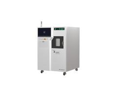
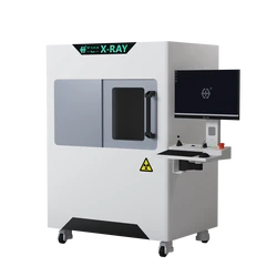
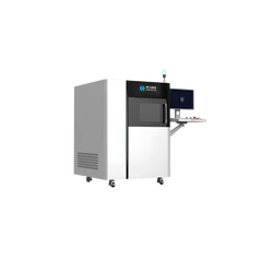
A confirmed Wahfei equipment image identifying the sample position and detector arrangement.