Durian X-Ray Inspection with 3D CT: See the Flesh Before the Fruit Is Opened
A durian is sold as a whole fruit, but what the buyer pays for is the flesh inside. 3D X-ray CT shows how much flesh a fruit actually carries — without cutting it open.
The value is hidden behind the husk
A durian's thick, spiky husk hides the part that matters. Inside, the flesh is arranged in several chambers, and how much flesh a fruit actually carries varies from one fruit to the next. The outside of the husk does not tell you.
In practice, grading still relies on experience: the shape of the fruit, its weight, the sound it makes when tapped. These are indirect signs, and even skilled graders disagree. Cutting the fruit open gives a definite answer — but a cut fruit must be sold immediately. The check itself destroys the product.
What the X-ray images show
With X-ray imaging, the fruit stays whole. The durian is placed on the rotary stage inside the cabinet (Figure 1). During scanning, the stage rotates the fruit through 360° while the detector captures a stream of projection images; a live transmission view is shown on the monitor throughout (Figure 2).
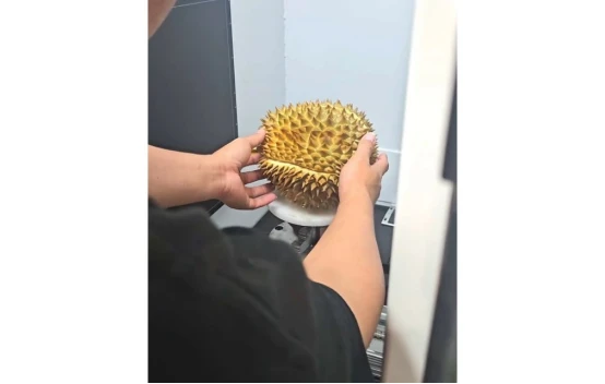
Figure 1 — A whole durian placed on the rotary stage of the HF-XT1000
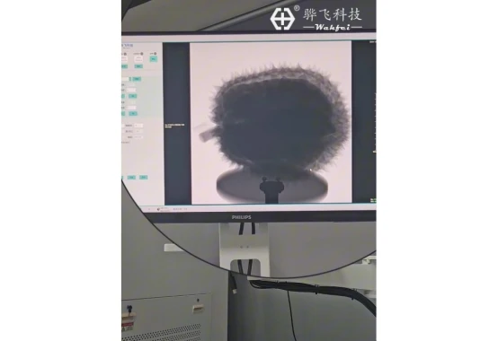
Figure 2 — Live transmission image of the fruit on the system monitor during scanning
The system reconstructs the scan into cross-sectional slices in real time. On a slice, the husk, the chambers and the flesh inside them become directly visible (Figure 3) — including how the flesh is distributed and how much of the fruit's volume it fills.
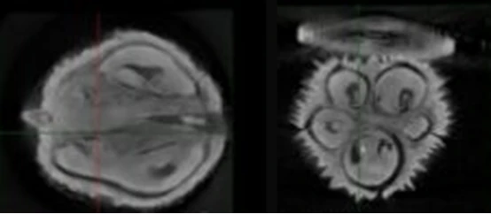
Figure 3 — Reconstructed cross-sectional slices: the chambers of flesh inside the husk are directly visible
This gives packers and exporters a practical basis for grading. Fruits can be sorted by flesh distribution and fill level before packing, and the scan data of each fruit can be saved for traceability. The fruit itself is untouched and remains saleable after scanning.
The system
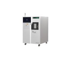
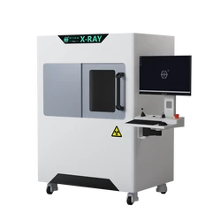
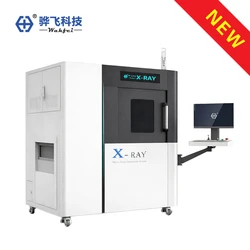
The images in this note were taken on the Wahfei HF-XT1000 3D X-ray CT inspection system (Figure 4), which combines a micro-focus sealed X-ray source with a high-resolution digital flat-panel detector, 360° rotation and real-time reconstruction in a single cabinet.
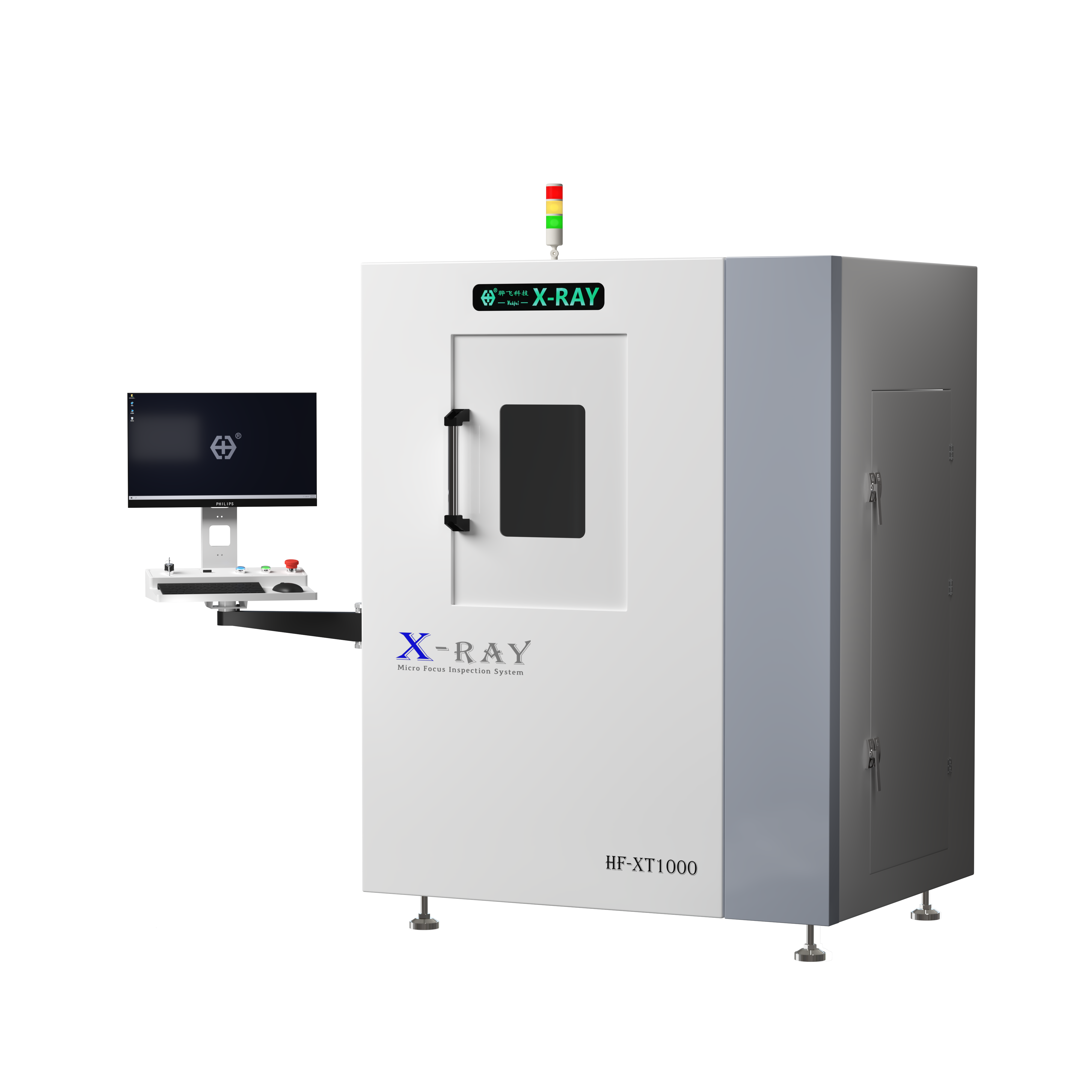
Figure 4 — The HF-XT1000 3D X-ray CT inspection system
For exporters, packers and distributors who handle durians in volume, non-destructive imaging turns grading from experience-based estimation into image-based evidence — and the fruit reaches the buyer intact.
About Wahfei Technology — Shenzhen Wahfei Technology Co.,Ltd. develops and manufactures industrial X-ray inspection systems. For HF-XT1000 specifications or a free sample evaluation, contact us at szwahfei@gmail.com or on WhatsApp at +86 189 0297 8624.
Note — The images are shown for illustration. Grading criteria for flesh distribution and fill level are defined by the buyer's own specification.